Table of Contents
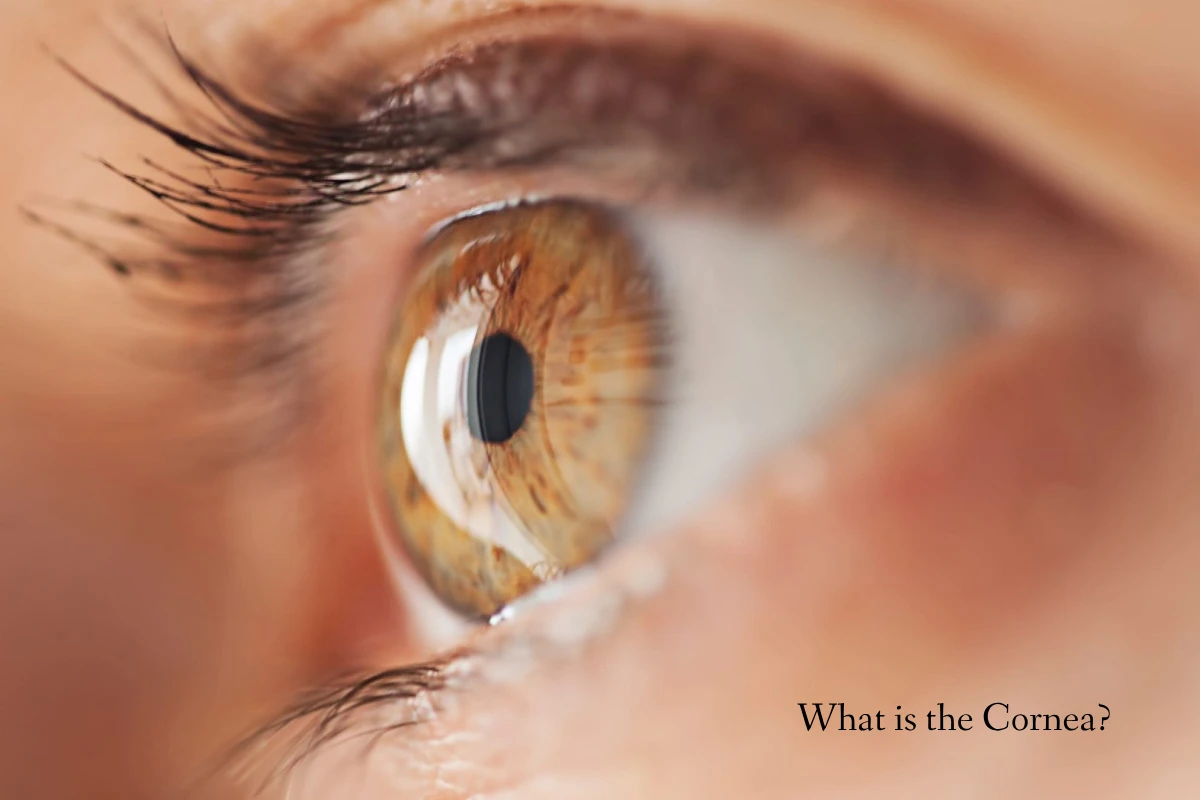
What is the Cornea?
The cornea is the outer layer of the eye. It is transparent, curvilinear and acts as the first lens that meets the light when it enters our eyeball.
For the correct function of the cornea, it must be kept transparent and must have an adequate curvature so that it fulfils good optical refractive properties.
The cornea and ocular surface are among the specialities that have received the most technical innovations in diagnosis and surgery in recent years.
Features of Cornea
For the anatomical and histological aspects of the tissue from which it makes, the cornea is:
Transparent: thanks to the geometry of the fabric and the absence of blood vessels.
Richly innervated: it is innervated by the upper branches of the fifth pair of Cranial Nerves, the trigeminal.
Mirror: reflects images of surrounding objects.
These characteristics allow it to be one of our natural lenses (dioptric means), the tools with which the eye processes information to send to the brain for correct vision.
Functions of the Cornea
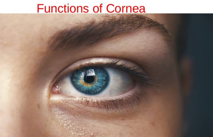
The cornea has three main functions, briefly described here.
1. The Mechanical Function
- The presence of collagen fibres and the very name of this organ (cornea means hard) account for its mechanical resistance characteristics.
- The mechanical properties of the cornea are necessary to maintain its shape over time, which is essential for preserving vision.
2. Support
- Thanks to its structure, the cornea allows the passage of nutritional molecules between the layers to make up for the absence of blood vessels.
3. Optics
- The cornea’s transparency allows the light rays to penetrate the eye and reach the retina, passing through the lens.
- It is the first of the dioptric means (the natural lenses of the eye) that light encounters.
- And also, its contribution to the convergence of images on the retina is equal to 65-75% of the total.
- By filtering some wavelengths of the UV spectrum, it allows light rays to pass through the fabric without being absorbed or reflected on its surface.
Layers of the Cornea
1. Corneal Epithelium
- 5/7 layers of cells from the corneal epithelium joined together and the basement membrane supported by anchoring fibres called hemidesmosomes.
- The function of the epithelium is to maintain the transparency of the cornea and protect it from external agents. And also, it has the property of rapid healing.
2. Bowman’s Stratum
- Bowman’s layer is a smooth, acellular, non-regenerative surface located between the eye’s epithelium, stroma, and cornea.
- As we age, this layer becomes thinner.
- Although the functionality of this membrane is still unclear, recent studies indicate that it acts as a physical barrier to protect the subepithelial nerve plexus and help sensory recovery and the innervation of the epithelium.
- It also helps to restore transparency. It is absent in non-primates such as cats, dogs and mice.
3. Corneal Stroma
- The stroma is the thickest part of the cornea in the eye. Its form of collagen fibres, approximately 200, arranged in sheets parallel to the surface, whose direction between them is oblique, helping the cornea’s transparency.
4. Cloak of Dua
- The Dua layer separates the last end of keratocytes in the cornea. And also, it is the final layer that forms the anatomy of the cornea.
5. Descemet’s Membrane
- Descemet’s membrane is made up of collagen IV fibres and, depending on age.
- This layer increases one micron in thickness every ten years.
- And also, its primary function is to serve as a modified basement membrane of the posterior epithelium or corneal endothelium.
6. Corneal Endothelium
- The corneal endothelium comprises hexagonal cells whose function is to dehydrate the cornea to keep it transparent.
- And also, as endothelial cells do not regenerate, they lost with age.
- In addition to age, that can occur due to some dystrophy, pathology or eye surgery.
- It can also happen that when there is a disproportionate number of cells, the cornea can lose its aqueous humour and, therefore, its transparency.
Causes of Cornea Damage
The most frequent are:
- Scratches on the cornea.
- Chemical injury: by harmful liquid that comes into contact with the eye.
- Abuse or misuse of contact lenses.
- Contact in the eye of external agents: the sensation of a foreign body (dust, sand);
- Excess ultraviolet rays in the eye.
- And also infections caused by fungi, viruses, or bacteria.
Furthermore, as mentioned above, the cornea’s transparency is one of the essential properties of the cornea. This transparency can be impaired when there is an absence of blood or lymphatic vessels, when it does not contain an adequate amount of proteoglycans (on which the regular ordering of stromal fibres depends) or when it has good hydration.
The sources to keep the cornea nourished are the capillaries of the limbus (they raise the peripheral part of the cornea), the tears (supply of glucose in the tear film).
And also, the aqueous humour (in charge of nourishing the outermost layers of the cornea, the endothelium being a unicellular layer).
Diseases of Cornea
There are many diseases of the cornea, both inherited and acquired. Due to their importance, we can highlight the following.
1. Corneal Opacity
- Corneal opacity is the loss of transparency of the cornea that can cause by trauma, inflammation, or hereditary causes.
- In these cases, a corneal transplant solves the problem.
2. Conjunctivitis
- Conjunctivitis is an infection that occurs in the conjunctiva, mucous membrane lining the inside of the eyelid.
- This can spread and also affect it, thus also compromising vision.
3. Blepharitis
- Blepharitis is inflammation of the eye rims caused by a malfunction of the Meibomian glands, responsible for lubricating the eye’s surface and preventing evaporation of the eye.
- And also, a watch with blepharitis implies inadequate hydration, therefore, of the cornea as these glands obstruct.
4. Corneal Ulcers
- The corneal ulcers involve, as the name itself, an ulcer in the outer layer of the eye, i.e. the cornea, usually caused by infection.
5. Keratitis
- Keratitis is a corneal inflammation whose causes can give dryness, viral infections, bacterial infections, or excessive sun exposure.
6. Dry Eye Syndrome
- Dry eye syndrome is the inability of the lacrimal system to maintain a lubricated and protected ocular surface. It manifests by a foreign body sensation, eye redness, tearing, and dry eyes.
- And also, it is prevalent in women in the 5th or 6th decades of life.
- There are many types of dry eye. Hence the success of the treatment depends on their correct identification.
7. Pterygium or Palm Tree
- The Pterygium or palm tree is the abnormal growth of the conjunctiva that can cover, making vision difficult.
8. Keratoconus
- The Keratoconus is particularly important in altering the structure of the cornea and thus distorts vision.
- This occurs because keratoconus is an alteration in the collagen fibres that make up the stroma.
- And, being weaker than average, the cornea deforms forward into a sphere shape.
9. Refractive Defects
- Refractive errors such as myopia, hyperopia, and astigmatism also occur in it.
- Nowadays, thanks to refractive surgery, it is possible to mould and change the shape of the cornea in the eye to dispense with glasses and contact lenses by improving its refractive state.
Conclusion
The cornea is a structure that is approximately in the shape of a dome that lines the eye’s anterior surface externally.